Anatomy Of Chest And Ribs - Normal female anatomy of the chest (thoracic) cavity and ... - And as you might guess from the word major, it makes up the majority of the chest muscle mass.
byAdmin•
0
Anatomy Of Chest And Ribs - Normal female anatomy of the chest (thoracic) cavity and ... - And as you might guess from the word major, it makes up the majority of the chest muscle mass.. Chest blunt trauma (cbt) and the resultant rib fractures often lead to thoracic collapse. The purpose of this study was to explore the effect of. Sternum, clavicles, scapulae, 12 pairs of ribs, 12 thoracic ve… protective framework for parts of the chest involved with brea… chest or thorax. The chest anatomy includes the pectoralis major, pectoralis minor and the serratus anterior. Anatomy is the amazing science.
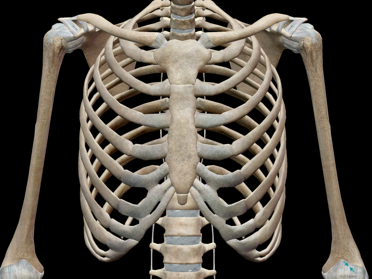
How these parts interrelate through joints is described also. It consists of the 12 thoracic vertebrae 12 pairs of ribs and the breastbone which is called the sternum. Major structures are shown in fig. O bones—spine, ribs, clavicles, scapulae, humeri. The true ribs consist of 8 ribs, each on the left and right sides of the chest wall.
3D Skeletal System: Bones of the Thoracic Cage from www.visiblebody.com Uppermost 10 pairs also articulate anteriorly with the sternum. ■ describe the anatomical relationships of various organs in the chest. Anatomy of the chest and the lungs: 5 centimeters far from tubercle, it suddenly changes its direction, this is termed angle of the rib. It discusses the specific anatomy of the ribs and costal cartilages, along with the sternum. Each rib wraps around the lung and descends approximately 3 to 5 inches. Anatomy is to physiology as geography is to history: They are ribbon like, elastic bony arches and flat in shape.
Moving during chest expansion to enable lung inflation.
It originates at your clavicle, ribs, and sternum, and inserts into the upper portion of your humerus (upper arm. It can help you understand our world more detailed and specific. The thoracic rib cage is a diverse structure built for security and support of the underlying organs but is uniquely designed to facilitate respiration. Paschalides medical publications, 2004, with. Continue scrolling to read more below. Basic rib anatomy consists of a head, neck, tubercle. ■ describe the anatomical relationships of various organs in the chest. The purpose of this study was to explore the effect of. Moving during chest expansion to enable lung inflation. Chest blunt trauma (cbt) and the resultant rib fractures often lead to thoracic collapse. It discusses the specific anatomy of the ribs and costal cartilages, along with the sternum. Pathology of the heart, mediastinum, lungs and pleura. Pectus excavatum is a congenital deformity of the ribs and the sternum producing a concave appearance of the anterior chest wall.
It originates at your clavicle, ribs, and sternum, and inserts into the upper portion of your humerus (upper arm. Each rib wraps around the lung and descends approximately 3 to 5 inches. Anatomy of the chest and the lungs: They are ribbon like, elastic bony arches and flat in shape. Describe the major bony features of the ribcage, the ribs and the thoracic vertebrae.
The Thoracic Cage | Anatomy and Physiology I from s3-us-west-2.amazonaws.com Describe the major bony features of the ribcage, the ribs and the thoracic vertebrae. Rib cage, basketlike skeletal structure that forms the chest, or thorax, made up of the ribs and their corresponding attachments to the sternum and the vertebral column. True, false and floating ribs are denoted. Identify the following structures on the lateral chest radiograph o diaphragm. ■ identify the basic anatomy seen on a chest radiograph. The purpose of this study was to explore the effect of. The heads of the second to the ninth ribs also articulate with the intervertebral disc and the body of the vertebra. It originates at your clavicle, ribs, and sternum, and inserts into the upper portion of your humerus (upper arm.
■ identify the basic anatomy seen on a chest radiograph.
Detailed anatomy of the rib cage | specific articulations. It can help you understand our world more detailed and specific. The ribs stretches posteriorly from thoracic vertebrae to the anterior lateral edges of the sternum. Basic rib anatomy consists of a head, neck, tubercle. Insert contains images of a typical rib and the first rib. This type of ct scan uses a lower radiation level than a conventional. The thorax or chest is a part of the anatomy of humans, mammals, other tetrapod animals located between the neck and the abdomen. Anatomy is the amazing science. The anatomy of a typical or a common rib. True, false and floating ribs are denoted. Describe the major bony features of the ribcage, the ribs and the thoracic vertebrae. Paschalides medical publications, 2004, with. Anatomy of the chest abdomen and pelvis was.
12 pairs of ribs which articulate posteriorly with the vertebral column. Continue scrolling to read more below. Pathology of the heart, mediastinum, lungs and pleura. As part of the bony thorax, the ribs protect the internal thoracic organs. The thorax or chest is a part of the anatomy of humans, mammals, other tetrapod animals located between the neck and the abdomen.
Interview with Marc Gosselin | Anatomy art, Skeleton ... from i.pinimg.com Anatomical illustrations this e anatomy module presents an illustrated anatomy of the lungs trachea bronchi pleural cavity and pulmonary ve. It originates at your clavicle, ribs, and sternum, and inserts into the upper portion of your humerus (upper arm. Detailed anatomy of the rib cage | specific articulations. Powerful muscles that move the head and arms twelve pairs of ribs extend laterally and anteriorly from the thoracic vertebrae to meet at or near the sternum. Chest the chest consists of bony skeleton of the spine and ribs, chest wall and diaphragm, the mediastinum and great vessels, the airways, lung posterior to the attachment of scalenus anterior, the subclavian artery and the lower trunk of the brachial plexus cross the rib and lie in contact with. It describes the theatre of events. It can help you understand our world more detailed and specific. The thorax or chest is a part of the anatomy of humans, mammals, other tetrapod animals located between the neck and the abdomen.
The purpose of this study was to explore the effect of.
The rib cage also anchors the bones of the head, neck, shoulders, and arms to the trunk of the body. 5 centimeters far from tubercle, it suddenly changes its direction, this is termed angle of the rib. Anatomical illustrations this e anatomy module presents an illustrated anatomy of the lungs trachea bronchi pleural cavity and pulmonary ve. The chest anatomy includes the pectoralis major, pectoralis minor and the serratus anterior. Paschalides medical publications, 2004, with. Chest blunt trauma (cbt) and the resultant rib fractures often lead to thoracic collapse. Continue scrolling to read more below. The bottom 2 pairs are referred to as 'floating ribs'. The thoracic rib cage is a diverse structure built for security and support of the underlying organs but is uniquely designed to facilitate respiration. Insert contains images of a typical rib and the first rib. O bones—spine, ribs, clavicles, scapulae, humeri. Side conclusion and anatomical position. Anatomy of the chest abdomen and pelvis was.
Side conclusion and anatomical position anatomy of chest. The ribs stretches posteriorly from thoracic vertebrae to the anterior lateral edges of the sternum.